Patient Going Strong Seven Years Past Brain Metastasis Diagnosis
Published
From the Duke Cancer Institute archives. Content may be out of date.
William Blake, 65, at his second home in the mountains of West Virginia
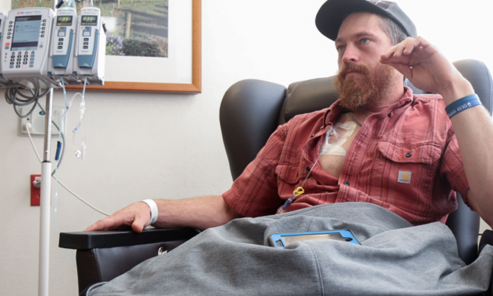
William Blake, 65, worked for the State of West Virginia for about 35 years in the toll division before taking early retirement in 2007 when he got sick with follicular lymphoma. He was treated at Charleston Area Medical Center (WV) and survived that cancer.
But when Blake was diagnosed four years later with a different type of cancer at the Beckley, WV, Veterans Hospital — metastatic melanoma to the brain — he was referred to Duke.
John Kirkpatrick, MD, PhD, the director of radiation oncology for The Duke Center for Brain and Spine Metastasis at Duke Cancer Institute and clinical director of radiation oncology at DCI, performed an image-guided stereotactic radiosurgery (SRS) procedure on Blake’s two brain lesions — a high-tech non-surgical therapy that delivers precisely-targeted high-dose radiation in one to five treatments, while preserving nearby healthy tissue.
Medical oncologist April Salama, MD, meanwhile, treated Blake with a course of ipilimumab, an immunotherapy that had just been approved that year for the treatment of advanced melanoma. When another lesion appeared in March 2012, Kirkpatrick “disappeared” that one as well.
“The prognosis was “We’ll do what we can, the mets are really small, and we caught it early,”” said Blake, who’s today making the most of his retirement.
While Blake has recently had some small localized melanomas and basal cell carcinomas removed in other areas of his body, there’s been no sign of cancer in his brain for seven years.
Brain metastasis survivor William Blake, 65, is enjoying his retirement, getting away on weekends to his cabin in the West Virginia mountains to read, hike, hunt, and fish, and spending more time with his wife and family and friends.
No Looking Back
Blake considers himself very fortunate. The percentage of patients who survive brain metastases beyond two years is traditionally less than five percent, but Duke is now seeing much better results than that. With treatment advances (primarily in surgery and radiosurgery) that offer excellent local control of brain metastases, coupled with lower rates of toxicity, an increasing number of patients are surviving well beyond that.
“We try to be realistic; I tell patients that there’s a lot of uncertainty with metastatic cancer, which is really one of the most difficult things in dealing with this disease, but that they should not look upon it as a death sentence,” said Kirkpatrick, who can recall a handful of patients with brain metastases who have lived more than a decade. “We don’t want to be Pollyanna, but on the other hand, you know, I’ve told enough people over the years that they will probably live six months to a year, and who very pleasantly surprise me by living a much longer period of time.”
After earning his doctorate in chemical engineering from Rice University in 1978, John Kirkpatrick, MD, PhD, made a career in Texas as an engineer and business manager in the chemical and plastics industry. But in 1995, he enrolled in medical school because he “wanted to do something that was really going to help people.” He found in radiation oncology a way to combine his engineering skills with day-in-day-out patient care. And he hasn’t looked back.
He explained that with each patient case he “plans for the worst and hopes for the best.” He’s seen the best, like William Blake’s case, and the worst.
Brain metastases can cause devastating symptoms that include headaches, seizures, vomiting, weakness, loss of vision, cognitive decline, and personality changes.
“We do our best to protect our patients’ brains by using the right surgery, the right radiation therapy, the right combination of drugs and the right supportive care so that those people are able to have a good quality of life and good cognition for as long as they can,” said Kirkpatrick.
Exponential Growth
Up to 30 to 40 percent of patients with solid tumors today can expect their cancer to metastasize to the brain. That’s because an increasing number of cancer patients are living longer due to better treatments — to the point where their primary cancer has a chance to spread to the brain.
Most common in patients with non-small cell lung cancer, breast cancer, and melanoma, brain metastases are also prevalent in patients with renal cell carcinoma (kidney cancer) and gastrointestinal cancers. However, any cancer can technically spread to the brain.
This rise in brain mets (metastasis) patients nationwide is driving more and more patients to seek treatment at Duke.
“The growth has been incredible. The number of patients being treated with stereotactic radiosurgery has grown 20 to 30 percent each year since 2002; from 10 patients in 2002 to more than 500 annually now,” said Kirkpatrick. “Our brain metastasis program is one of the biggest programs of its kind in the country. Our team of physicians, physicists, radiation therapists, dosimetrists and nurses does a very good job of treating patients really well when they come here — using the best available technology in surgery and radiotherapy — and in some cases bringing new anti-cancer drugs into the picture as well.”
Results from a new clinical trial, recently published in JAMA Surgery, suggest that a gene-based assay could help physicians better assess the risk of cancer spread in patients with early-stage melanoma, potentially sparing some patients from unnecessary surgery.The study, which included investigators from the Duke Cancer Institute (DCI), is the first prospective clinical trial to evaluate whether a molecular gene signature can improve risk stratification for patients whose melanoma is otherwise considered early-stage based on traditional staging methods.Melanoma continues to rise in incidence, and most patients are diagnosed at an early stage of disease. Standard melanoma staging relies on the tumor, node, metastasis (TNM) system, which classifies tumors based on size, lymph node involvement, and whether the cancer has spread to distant organs.While TNM staging is useful, it does not always capture how aggressive a tumor may be. Some patients diagnosed with stage I melanoma still experience disease recurrence or lymph node spread, outcomes that are not fully explained by traditional clinical and pathologic features alone.“By relying only on conventional staging, we may be missing some of the ‘bad actors,’” said Georgia Beasley, MD, DCI surgical oncologist and co-investigator on the trial. “Other cancers, like breast cancer, have increasingly incorporated gene-based tools to help better predict risk, and melanoma has lagged behind in that regard.”The trial evaluated a gene expression assay known as the Merlin test, which analyzes tumor tissue from a patient’s initial melanoma biopsy. The assay combines genetic information from the tumor with established clinical features to generate a personalized risk estimate for lymph node spread.Unlike previous studies, which examined gene signatures retrospectively, this was the first large, prospective clinical trial to test whether such an approach could meaningfully add to existing risk prediction for early-stage melanoma patients.One of the most immediate potential benefits of the test is its ability to help guide surgical decision-making, particularly regarding whether a patient should undergo sentinel lymph node biopsy, a procedure that requires anesthesia and carries some risk of complications.This consideration is especially relevant for older patients or those with additional medical conditions.“If we have a tool that tells us a patient’s likelihood of lymph node involvement is very low, we may be able to safely avoid more aggressive surgery,” Beasley said. “That means avoiding anesthesia, reducing complications, and overall lowering the burden of treatment.”For both patients and physicians, the test may help reduce uncertainty. While many patients understandably want to pursue every available intervention, having a reliable, personalized risk estimate could make it easier to choose less invasive options when appropriate.Following publication of the trial results, the Merlin assay is now commercially available and can be ordered by physicians, including those at Duke, for selected patients.Beasley noted that this represents an important step toward integrating molecular risk tools into everyday melanoma care. However, she emphasized that further refinements are needed to improve efficiency, particularly in how tumor samples are handled.“Right now, the process still involves shipping glass pathology slides, which can slow things down,” Beasley said. “Ideally, this would all move to a digital platform so results could be generated faster and patients wouldn’t have to wait weeks for answers.”Work is already underway to streamline and digitize parts of this process, with the goal of delivering timely information that can better inform real-time treatment decisions.While biomarkers and risk assessment tools may not draw the same attention as novel cancer drugs, Beasley emphasized that their impact on patient care can be substantial.“These kinds of studies are incredibly important, even if they’re not flashy,” she said. “They help patients, families, and physicians make better decisions, and that’s something that’s often underestimated.”