From the Duke Cancer Institute archives. Content may be out of date.
Walks in the park, a stress-relieving game of fetch, affectionate belly rubs. “Man’s best friend” brings us all that and more.
But, in the case of bone cancer (osteosarcoma), dogs are proving, again, that they can benefit their humans’ lives in a substantial way. Duke orthopedic oncologist Will Eward, MD, DVM, and colleagues are investigating how this link could improve health outcomes for children stricken by this disease.
The Case for Osteosarcoma and Dogs
Duke oncologist and veterinarian Will Eward with veterinary oncologist Steven Suter and cancer patient Deuce.
Osteosarcoma rarely occurs in humans, accounting for less than one percent of all diagnosed cancers. However, when it appears, it impacts children most heavily as the third most commonly occurring pediatric cancer after lymphoma and brain cancer.
Treating it is difficult, though, because there’s been lack of therapeutic advancements. “With osteosarcoma, I’m telling parents exactly what was told to them in the 1980s,” Eward said. “We can’t keep driving the 1984 Datsun forever. We have to do better because pediatric oncology occupies a special place in medicine. Kids shouldn’t get cancer.”
The small number of children developing osteosarcoma has been a barrier to improving care. That’s where dogs come in, he says. Veterinarians diagnose roughly 4 million dogs with cancer annually, and osteosarcoma accounts for approximately 15 percent of those cases. Not only are most of their tumors identical to those in humans, but their immune systems are also similar, and they share the same food, air, and water exposures. Additionally, the natural survival time for a dog with osteosarcoma is approximately 18 months, making it quicker to find out if treatments prolong life.
To test the viability of new drugs, Eward is partnering, via the Consortium for Canine Comparative Oncology, with veterinary oncologist, Steven Suter, VMD, PhD, medical director of North Carolina State College of Veterinary Medicine’s canine/feline molecular oncology diagnostic lab. Through a canine clinical trial, funded by a $250,000 grant from Hyundai Hope on Wheels to specifically target links between canine and human sarcomas, they are investigating whether two existing medications can combat osteosarcoma in dogs, potentially opening the door for use with children.
“These are dogs that just happen to get osteosarcoma like we do,” Eward says. “This is a clinical trial for dogs just like it would be for humans. Their owners have asked for their pets that have osteosarcoma to be included, and we’re treating them the same as we would in a human clinical trial even though that group has two legs and this group has four legs and wags their tails.”
Testing Therapies
A drawing of a low-density lipoprotein--a naturally produced substance in the body that cancers eat. Duke engineer David Needham used nanotechnology to encapsulate an existing drug into a tiny particle that mimics low-density lipoprotein.
The clinical trials test two existing medications approved for other uses—bortezomib and niclosamide. The team designed these trials based on predictive work conducted in mice.
Using immunosuppressed mice, researchers Jason Somarelli, PhD, and David Hsu, MD, PhD, created dog avatars, called xenografts, by injecting cells from dog tumors into the mice to prompt tumor growth. Doing so allowed researchers to more quickly see the drug’s effect.
“Using these xenografts, we demonstrated on a small scale that the drugs could work and could be scaled up for use from a 30-gram mouse to a 150-pound dog,” Somarelli says.
“This type of collaboration between bench science and veterinarians is critical because you can reach discovery much faster and at less cost. We haven’t spent time or millions of dollars going down the rabbit hole," he adds.
In the first trial, after seeing strong efficacy with the xenografts, the team examined how well bortezomib, a drug used to treat multiple myeloma cancer in humans, would work in dogs. While other chemotherapy agents attack cells indiscriminately, causing toxic side effects, bortezomib avoids those responses by only targeting how the cancer packages proteins. Consequently, this trial enrolled 10 dogs who received standard amputation but whose owners did not want to pursue subsequent chemotherapy.
Despite high expectations, Suter says, bortezomib has performed poorly, introducing neurological side effects, such as limb weakness and an inability for the dogs to walk. In addition, the dogs who received the drug didn’t fare any better than those who received standard chemotherapy. While it’s disappointing that bortezomib doesn’t work as anticipated, Eward says, the trial results are still critical.
“If we’ve found that bortezomib has no role for treating osteosarcoma, we’ve figured that out by treating 10 dogs for $90,000 in one year rather than treating five to 10 children in a trial that cost over $1 million,” he said. “We’ve saved doctors from using something that’s not effective.”
The second trial is focused on niclosamide, a drug traditionally used to treat intestinal parasites. Although existing evidence revealed it had some efficacy against osteosarcoma, as an oral drug, it wasn’t a viable treatment option because it can’t dissolve in the blood to reach the tumors. So David Needham, Ph.D., Duke professor of mechanical engineering and material science, created a version of niclosamide that could be intravenously administered, called niclosamide stearate pro-drug therapeutic (NSPT). He used nanotechnology to encapsulate niclosamide into a particle that mimics low-density lipoprotein (a naturally produced substance in the body that cancers eat).
Ten enrolled dogs have received amputation, four standard chemotherapy treatments, and four doses of the modified niclosamide.
To date, Suter says, the results have been promising, with no organ toxicity, no neurological impacts, and only some allergic reactions that can be treated with antihistamines.
The Importance of Collaboration
Researchers Jason Somarelli, PhD, and So Young Kim, PhD.
Ultimately, Eward says, the partnership between Duke and N.C. State has been crucial in moving osteosarcoma research forward. In emerging from their individual silos, bench scientists, oncologists, and veterinarians are charting new territory in cancer treatment. Much work still needs to be done in determining how NSPT kills cancer, providing the therapy to more dogs, and testing NSPT in other solid tumors, but it is clear the potential exists for this drug to combat additional cancers, including breast, prostate, and pancreatic.
“This is kind of like A Tale of Two Cities, only it’s a tale of two drugs,” Eward says. “We had two promising therapies based on the results of mice trials, and now we’re putting effort into moving NSPT to the next step to, one day, be available to humans with this disease.”
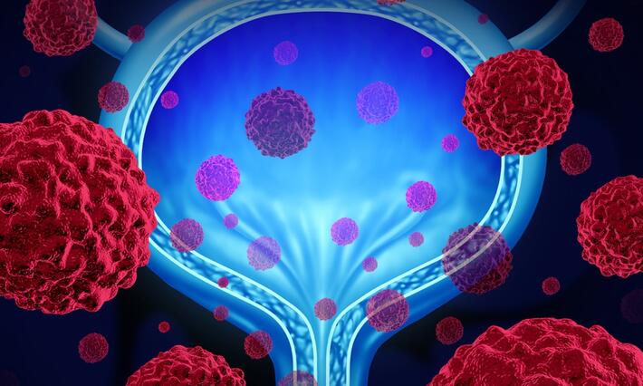
New research from Duke Cancer Institute (DCI) is helping uncover why some bladder cancer patients are less likely to benefit from a common treatment and how a patient’s cancer history may help guide more personalized care decisions in the future.For more than 50 years, Bacillus Calmette-Guérin (BCG) treatment has been the standard of care for patients with early-stage bladder cancer. It helps reduce the chances that cancer will come back after surgery, and it works well for many patients. But not all bladder cancers behave the same.Bladder cancer is one form of a broader group known as urothelial cancers, which can develop anywhere along the urinary tract. Most cases begin in the bladder itself and are often caught early because patients develop symptoms, such as blood in the urine. These early-stage cancers are typically treated with surgery followed by BCG, which is delivered directly into the bladder to help prevent recurrence.However, about 10 percent of urothelial cancers originate in the upper urinary tract—in the kidneys or ureters. Even after those tumors are treated, cancer can later reappear in the bladder.“Historically, we’ve treated these bladder recurrences the same way we treat primary bladder cancer,” said Yu Guang Tan, MD, a urology fellow at Duke and a collaborator from Duke-NUS Medical School in Singapore. “But the question is, are they really the same disease?”In a recent study published in Urologic Oncology, Tan worked with colleagues including Michael Abern, MD, co-chair of the DCI Center for Prostate and Urologic Cancers, to explore whether bladder cancers that arise after upper tract disease respond differently to BCG than cancers that originate in the bladder.Across both a single-institution study and a larger systematic review and meta-analysis of more than 1,300 patients worldwide, the team found a consistent pattern: patients with a history of upper tract urothelial cancer were significantly more likely to experience recurrence or disease progression after BCG treatment.“In some cases, nearly half of these patients did not respond well to BCG,” Tan said. “That’s a substantial number, and it suggests we may need to think differently about how we treat this group.”BCG has long been the backbone of therapy for non-muscle invasive bladder cancer, but it is not without limitations. In addition to variable effectiveness, the treatment has also faced periodic shortages, particularly in smaller or rural care settings.“This research gives us another piece of the puzzle,” Abern said. “If a patient has a history of upper tract disease, it may shape expectations about how well BCG will work and whether we should consider other options sooner, including clinical trials.”Tan and Abern both emphasize that BCG remains an important and effective therapy for many patients. The goal is not to replace it, but to better match treatments to the patients most likely to benefit.One of the most important implications of the study is its potential role in advancing precision medicine for bladder cancer. The team found that while these cancers may look similar under the microscope, they are likely biologically distinct. That difference may explain why they respond differently to treatment.“We think this history of upper tract cancer may act as a clinical biomarker,” Tan said. “It helps us predict how a patient might respond, but we still need to understand the biology behind it.”To answer that question, the researchers are now conducting whole genome sequencing studies to identify the genetic differences between these tumor types. They are also building a joint database between Duke and Duke-NUS to uncover additional factors that may influence treatment outcomes.“It’s an exciting time in bladder cancer research,” Tan said. “We have many new therapies emerging, and the challenge now is figuring out which treatment is best for which patient.”The research reflects a strong international partnership between Duke University and Duke-NUS Medical School in Singapore. By bringing together data from multiple countries and healthcare systems, the team was able to confirm that these findings are consistent across diverse patient populations.“That global perspective is really important,” Abern said. “It shows that this trend holds true across different regions, which makes the findings more robust and meaningful.”As new therapies, including immunotherapy and targeted treatments, continue to emerge, studies like this one will play a key role in shaping how patients are selected for different treatment approaches.“By better understanding these differences, we can improve how we counsel patients, design clinical trials, and ultimately deliver more personalized care,” Abern said.
New research from Duke Cancer Institute (DCI) is helping uncover why some bladder cancer patients are less likely to benefit from a common treatment and how a patient’s cancer history may help guide more personalized care decisions in the future.For more than 50 years, Bacillus Calmette-Guérin (BCG) treatment has been the standard of care for patients with early-stage bladder cancer. It helps reduce the chances that cancer will come back after surgery, and it works well for many patients. But not all bladder cancers behave the same.Bladder cancer is one form of a broader group known as urothelial cancers, which can develop anywhere along the urinary tract. Most cases begin in the bladder itself and are often caught early because patients develop symptoms, such as blood in the urine. These early-stage cancers are typically treated with surgery followed by BCG, which is delivered directly into the bladder to help prevent recurrence.However, about 10 percent of urothelial cancers originate in the upper urinary tract—in the kidneys or ureters. Even after those tumors are treated, cancer can later reappear in the bladder.“Historically, we’ve treated these bladder recurrences the same way we treat primary bladder cancer,” said Yu Guang Tan, MD, a urology fellow at Duke and a collaborator from Duke-NUS Medical School in Singapore. “But the question is, are they really the same disease?”In a recent study published in Urologic Oncology, Tan worked with colleagues including Michael Abern, MD, co-chair of the DCI Center for Prostate and Urologic Cancers, to explore whether bladder cancers that arise after upper tract disease respond differently to BCG than cancers that originate in the bladder.Across both a single-institution study and a larger systematic review and meta-analysis of more than 1,300 patients worldwide, the team found a consistent pattern: patients with a history of upper tract urothelial cancer were significantly more likely to experience recurrence or disease progression after BCG treatment.“In some cases, nearly half of these patients did not respond well to BCG,” Tan said. “That’s a substantial number, and it suggests we may need to think differently about how we treat this group.”BCG has long been the backbone of therapy for non-muscle invasive bladder cancer, but it is not without limitations. In addition to variable effectiveness, the treatment has also faced periodic shortages, particularly in smaller or rural care settings.“This research gives us another piece of the puzzle,” Abern said. “If a patient has a history of upper tract disease, it may shape expectations about how well BCG will work and whether we should consider other options sooner, including clinical trials.”Tan and Abern both emphasize that BCG remains an important and effective therapy for many patients. The goal is not to replace it, but to better match treatments to the patients most likely to benefit.One of the most important implications of the study is its potential role in advancing precision medicine for bladder cancer. The team found that while these cancers may look similar under the microscope, they are likely biologically distinct. That difference may explain why they respond differently to treatment.“We think this history of upper tract cancer may act as a clinical biomarker,” Tan said. “It helps us predict how a patient might respond, but we still need to understand the biology behind it.”To answer that question, the researchers are now conducting whole genome sequencing studies to identify the genetic differences between these tumor types. They are also building a joint database between Duke and Duke-NUS to uncover additional factors that may influence treatment outcomes.“It’s an exciting time in bladder cancer research,” Tan said. “We have many new therapies emerging, and the challenge now is figuring out which treatment is best for which patient.”The research reflects a strong international partnership between Duke University and Duke-NUS Medical School in Singapore. By bringing together data from multiple countries and healthcare systems, the team was able to confirm that these findings are consistent across diverse patient populations.“That global perspective is really important,” Abern said. “It shows that this trend holds true across different regions, which makes the findings more robust and meaningful.”As new therapies, including immunotherapy and targeted treatments, continue to emerge, studies like this one will play a key role in shaping how patients are selected for different treatment approaches.“By better understanding these differences, we can improve how we counsel patients, design clinical trials, and ultimately deliver more personalized care,” Abern said.